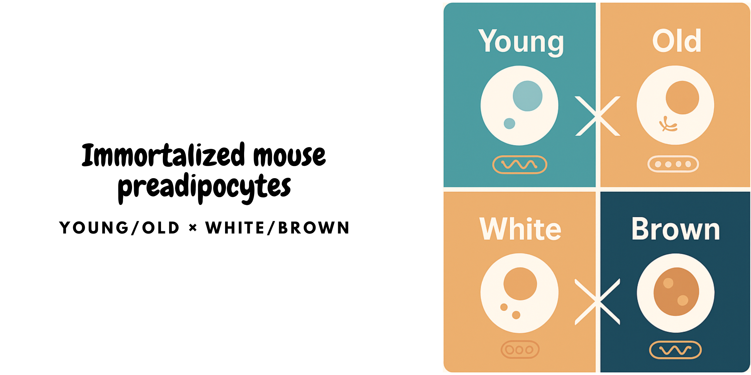
New Immortalized Mouse Preadipocyte Cell Lines from UMD Now Available

We’re excited to offer four research-ready cell lines engineered by Dr. Sui Seng Tee’s team at the University of Maryland School of Medicine (UMD). These immortalized preadipocytes enable consistent, reproducible studies of adipogenesis, thermogenesis, aging, metabolic disease, and drug response — without repeated animal isolations.
Immortalized Mouse Preadipocyte Cell Lines — Young & Old • Brown & White
Browse the four cell linesWhy these lines
- Balanced panel:
- Protocol-aligned:
- Consistency:
- Cryo-ready:
- Adipogenesis & thermogenesis assays (e.g., β-adrenergic activation)
- Aging comparisons: nuclear size, mitochondrial morphology, lipid droplet metrics
- Metabolic disease modeling (obesity, insulin resistance, NAFLD)
- Compound screening & nutraceutical evaluation
See protocol-aligned tips in the FAQs below.
Shop the Panel
Immortalized Young Mouse Brown Preadipocyte Cell Line
Derived from interscapular brown adipose tissue (iBAT) of 1-day-old pups; ideal baseline for thermogenic studies.
View productImmortalized Old Mouse Brown Preadipocyte Cell Line
Established from 27-month-old mice; supports aging comparisons in brown adipose biology.
View productImmortalized Young Mouse White Preadipocyte Cell Line
Subcutaneous WAT control to pair with young brown; useful for adipokine and lipid metabolism studies.
View productImmortalized Old Mouse White Preadipocyte Cell Line
Aged sWAT line for senescence, SASP, and inflammation readouts in white adipocytes.
View productFrequently Asked Questions
How fast do these lines differentiate?
Brown preadipocytes typically reach mature adipocytes in ~12 days under induction and maintenance media with insulin, IBMX, indomethacin, dexamethasone, rosiglitazone, and T3. White lines typically mature in 8–10 days without T3.
What markers are supported?
Standard readouts include UCP-1 (brown), PPARγ and FABP4 (brown & white), plus morphology-based lipid droplet metrics and mitochondrial staining (e.g., MitoTracker).
Any aging phenotypes I can compare?
Yes. Quantify nuclear size distributions, mitochondrial network integrity, and lipid droplet size distributions across young vs. old cohorts to model age-associated remodeling.
Culture & handling pointers
Use DMEM/F-12 + 10% FBS for growth; pellet cells gently (~600 g, ~3 min). Use polybrene during retroviral transduction in protocols where applicable; optimize concentrations for your lab setup.
Reference & Acknowledgment
This panel and usage guidance are aligned with protocols and data reported by Dr. Sui Seng Tee and colleagues (University of Maryland School of Medicine), detailing establishment, immortalization, selection, cryopreservation, and differentiation of mouse brown and white preadipocytes from young and aged animals, including marker validation (UCP-1, PPARγ, FABP4), lipid droplet imaging, and mitochondrial morphology analyses.
 Loading ....
